Amena Ali M Al-Sakran1,
Promy Virk1 ![]() ,
Mai Elobeid1,
Sherifa Shaker Hamed1,3,
Muzammil Igbal Siddiqui1,
Sawsan Omer4,
Nada Mohammed Mirghani2
,
Mai Elobeid1,
Sherifa Shaker Hamed1,3,
Muzammil Igbal Siddiqui1,
Sawsan Omer4,
Nada Mohammed Mirghani2
For correspondence:- Promy Virk Email: virkg@hotmail.com Tel:+96610502205785
Received: 15 May 2015 Accepted: 29 November 2015 Published: 29 January 2016
Citation: Al-Sakran AA, Virk P, Elobeid M, Hamed SS, Siddiqui MI, Omer S, et al. Histopathological effects on testis of adult male carp, Cyprinus carpio carpio, following exposure to graded concentrations of water-borne bisphenol A. Trop J Pharm Res 2016; 15(1):73-80 doi: 10.4314/tjpr.v15i1.10
© 2016 The authors.
This is an Open Access article that uses a funding model which does not charge readers or their institutions for access and distributed under the terms of the Creative Commons Attribution License (http://creativecommons.org/licenses/by/4.0) and the Budapest Open Access Initiative (http://www.budapestopenaccessinitiative.org/read), which permit unrestricted use, distribution, and reproduction in any medium, provided the original work is properly credited..
Purpose: To evaluate the estrogenic effect of Bisphenol A (BPA), an endocrine disruptor on the histological features in carp testis
Methods: Adult male fish, koi carp, Cyprinus carpio carpio, were exposed to three graded concentrations of BPA (10, 100 and 1000 µg/L) for a period of 21 days. A single dose of 17-β estradiol (1 ng/L) was used as positive control. The end points assessed at the end of the exposure period were condition factor, hepatosomatic index (HSI), gonadosomatic index (GSI), histopathological changes in the testis and lobular diameter.
Results: BPA caused a significant decrease in gonadosomatic index (GSI) of the fish at the median concentration of 100 µg/L. The major alterations observed in the gonad structure were a significant decrease (p ≤ 0.001) in the lobular diameter (65.1 ± 12.2 µm) compared with control (211.7 ± 36.60 µm) and complete loss in lobular structure with degenerating spermatozoa in some carps. The histopathological effects also include delayed sperm maturation and impaired spermatogenesis.
Conclusion: The findings clearly show marked adverse histopathological effects of gonads of adult carps when exposed to BPA.
Introduction
Xenoestrogens are endocrine disrupting chemicals (EDCs) that mimic the natural estrogens and have been reported to cause endocrine disarray in fish and other aquatic organisms [1]. Of all such xenoestrogens registered for use by the Environmental Protection Agency (EPA), the monomer, BPA (2,2-bis-(4-hydroxyphenyl)- propane, an alkyl phenol has perhaps triggered the greatest amount of interest among researchers and its use has been a debatable issue during the past decade[2]. While the point sources of BPA in the environment are sewage effluent and landfill leachate, fragments of epoxy resins and polycarbonate plastic debris entering the watershed through runoff make up the non-point sources [3]. The ubiquity of BPA in the aquatic environment is well established [4], with BPA accumulating in water systems from anthropogenic sources [2].
The estrogenic potency of BPA is well documented [2,5-8]. Effects of endocrine disrupting chemicals like BPA on fish are evaluated by gross gonad morphology and histology [6,7]. Since exogenous estrogens have the potential to interfere with the sexual development and reproduction of aquatic vertebrates [9], reproductive ability is a sensitive indicator of stress induced by these compounds. Thus an evaluation of reproductive dysfunction based on gonad morphology and spermatogenic endpoints are ideal biomarkers.
The present study was aimed at assessing and understanding the deleterious alterations in the testes of sexually mature cyprinid, Koi carp Cyprinus carpio carpio exposed to graded concentrations of Bisphenol A (BPA) for 21 days. The key endpoints assessed at the end of the exposure period were the somatic parameters and histopathology of the testis.
Methods
Preparation of fish
One year-old mature male koi carp Cyprinus carpio carpio (n = 60, mean body mass = 150-200 g, and mean body length = 22 cm) were obtained from a local fish farm near Riyadh city in October 2012 and were divided into six exposure groups of 10 fish each. Prior to the experimental period the fish were acclimatized to laboratory conditions in glass aquaria for 15 days. During this period, each glass aquarium was filled with 100 L of aerated and de-chlorinated tap water. Natural 12 h light: 12 h dark schedule was maintained. Temperature was maintained between 24 - 26 oC and dissolved oxygen was at least 90 % saturation at all times. During the acclimatization and exposure period, fish were fed a commercial diet of dry pellet feed at 1 % body weight per day. All animal procedures were performed in accordance with the standards set forth in the Guidelines for the Care and Use of Experimental Animals by the Committee for the Purpose of Control and Supervision of Experiments on Animals (CPCSEA) [10]. The study protocol was approved by the Animal Ethics Committee of the Zoology Department, College of Science of King Saud University, Saudi Arabia (approval no. 799432).
BPA exposure
Following acclimatization the fish were exposed to different water-borne concentrations of BPA (10, 100, 1000 µg/L) for 21 days. Analytical grade BPA (CAS Registry no. 80-05-7, 98 %, Lobachemie, India) was used as the test xenoestrogen and 17β-Estradiol (E2) (97 %, Lobachemie, India) was used as a positive control. Stock solutions of both the chemicals were prepared in acetone (99.5 %, Schartab S.L, Spain).The stock solutions were used to prepare the three dosing test solutions with nominal concentrations of BPA (10, 100, 1000 µg/L) and one test solution with nominal concentration of E2 (1 ng/L). The two remaining glass aquarium were negative controls, one receiving only dechlorinated tap water while the other was the vehicle (acetone) as control.
During the exposure period, the water, BPA, E2 and acetone of each glass aquarium were changed after 7 days and the physico-chemical parameters of water were analyzed at regular intervals and were in the conducive range. There was no mortality during the exposure period. After 21 days of exposure seven fish from each group were taken out and anaesthetized. Body mass (nearest gram) and the body length (nearest mm) of each fish was recorded. The fish were subsequently sacrificed, the liver and gonads excised and weighed separately to assess the hepatosomatic index (HSI) and gonadosomatic index (GSI). Condition Factor (K), HSI and GSI were calculated as in Eqs 1 – 3, respectively [11-12]:
K = W/L3 ……………………………….. (1)
where W = weight of fish and L = length of fish.
HSI = (Lm/Bm)100) ………………...…. (2)
GSI =/[Gm/Bm)100…………………….. (3)
where Gm is gonad mass.
Histological techniques and photomicro-graphy
Samples of fragments of fresh testes were fixed in 10 % buffered formalin. Sections of the testes were taken from the apical portion for all groups. Thereafter the tissues were dehydrated through a graded series of ethanol (from 70 to 100 % ethanol in subsequent steps). Xylene was used as a clearing agent. Tissues were embedded in paraffin (58.6 ºC). Sections (5 µm) were stained with haematoxylin and eosin, and were examined and photographed using a photomicroscope. Stages of spermatogenesis were determined according to the germ cell types present and their relative abundance [13]. The histopathological alterations were assessed on reduction of average lobule diameter and alterations in the germ cell types.
Determination of lobular diameter
The average diameter of the seminiferous lobules was measured using a calibrated ocular scale by randomly selecting twenty lobules of comparable shape per gonad.
Statistical analysis
Since the data in control and solvent control showed no significant difference, they were pooled together. All presented data are expressed as mean values ± standard deviation (SD). Group differences were analyzed with unpaired Student’s t-test. Numerical data was correlated with SPSS 16.0 statistical software (Chicago, IL, USA). The level of significance was set at p ≤ 0.05.
Results
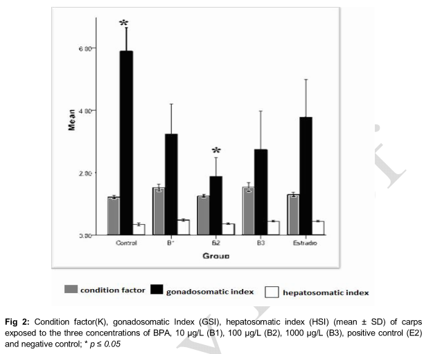
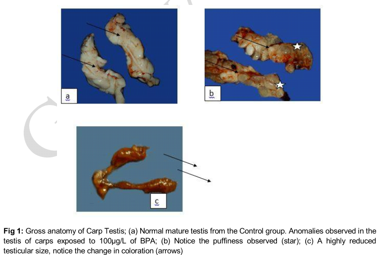
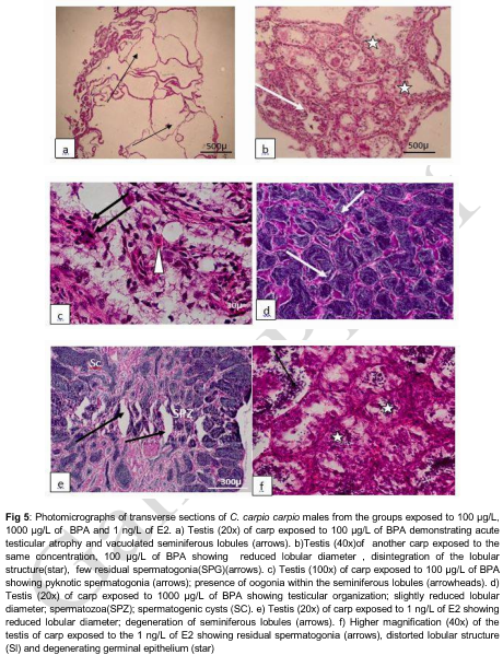
Exposure to the BPA and the E2 at levels employed in the present study did not cause any mortality nor did it affect the growth of the fish as there was no significant difference in the condition factor of fish from all the experimental groups post exposure (). Distinct anatomical anomalies were observed in the testes of carps exposed to 100 µg/L of BPA ( b,c) in comparison to those from the control group (a).
Marked histopathological alterations were observed in the testes of carps exposed to the low doses of BPA (10 and 100 µg/L) and E2 (positive control) in comparison to the control group.
Somatic parameters
There was no significant difference observed in the Condition factor and Hepatosomatic Index of carps in the control and the treated groups. However, a significant decrease (p ≤ 0.01) in the GSI was observed in the group exposed to 100 µg/L of BPA (1.8) in comparison to the control (5.9). The GSI of carps in other treated groups did not vary significantly from the control ().
Diameter of seminiferous lobules
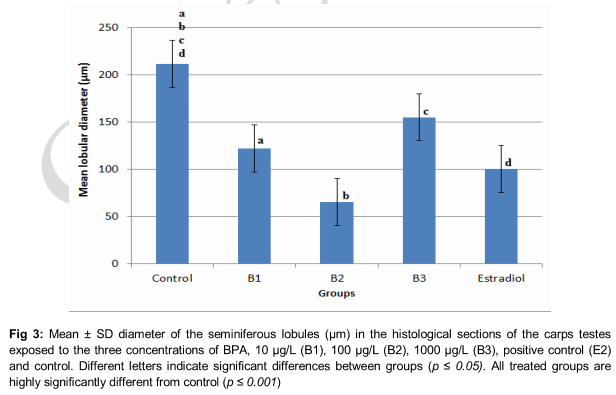
Testes of carps in all the treated groups showed a statistically significant (p ≤ 0.001) decrease in the lobular diameter in comparison to the control (211.7 ± 36.59 µm) being marked in the group exposed to 100 µg/L BPA (65.1 ± 12.15 µm) followed by the positive control group exposed to 1 ng/L E2 (100.6 ± 17.77 µm) ().
Histopathology of testis
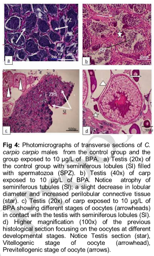
The testes of the control fish showed the typical organization of the carp testis with large seminiferous lobules (SL) and the interlobular spaces with interstitial cells and blood vessels. The two stages of maturity exhibited in the testis were mainly the spermiation stage, with free spermatozoa (SPZ) filled in the lobular lumen and active spermatogenesis characterized by spermatogenic cysts (SC) ( a).
Starting from the lowest concentration of 10 µg/L of BPA, the exposed fish showed a decreased lobular diameter (). Although there was noalteration in the lobular structure a decreased intensity in the mature free SPZ in the lobular lumen was observed in the sections of testes of carps exposed to10 µg/L of BPA. This reflected a delayed maturation as more number of spermatogonia (SPG) and spermatocytes (SPC) were observed in the lobules (b). A few sections of the carp testis exposed to the same concentration showed the presence of developmental stages of oocytes associated with the testicular tissue (c and d). At a higher concentration of 100 µg/L pronounced testicular atrophy was observed in some exposed fish. This was characterized by a loss of lobular organization, presence of lacunae with disintegration of the germinal epithelium. A few distorted lobules showed residual germ cells and SPG (a). Other carps exposed to the same concentration (100 µg/L) showed a significantly reduced lobular size with lacunae and SPG with a few primary oocytes (Figs. 5 b and c). Overall, the effect observed indicates inhibition of spermatogenesis and loss of testicular organization.
At the highest concentration of BPA (1000 µg/L), the exposed fish did not show any alteration in the lobular structure. The testicular organization was intact comparable to the control with advanced stage of spermatogenesis characterized by the presence of SC and SPZ. However the lobular diameter was reduced (d). The testis of the carps exposed to 1 ng/L E2 (positive control) showed a regressed testis with diminished lobular size. Spermatogenesis was inhibited with residual SPZ in the lobular lumen. The lobules had vacuolar spaces with increased amorphous matrix in the interlobular space. The effects are comparable to the most affected group of carps exposed to 100 µg/L of BPA (Figs 5 e and f).
Discussion
The endocrine disruptive nature of BPA which impairs reproductive physiology has been extensively reviewed in the past based on comparative studies both in mammals and fish [8]. There is a great deal of evidence that in vivo experimental exposure of adult male fish to BPA has detrimental effects on the gonadal morphology and histology. A few studies that can be cited with their findings are decreased spermatozoa and abnormal testicular connective tissue in Japanese medaka Oryzias latipes [15], reduced GSI and inhibition of spermatogenesis in fathead minnow Pimephales promelas [6], loss of testicular structure and increase in fibrotic tissue and decrease in number of spermatozoa in Japanese medaka Oryzias latipes [16], adverse effect on sperm quality in brown trout Salmo trutta fario [17], impaired steroidogenesis and altered gonad histology in adult common carp Cyprinus carpio [7], and impaired steroidogenesis and sperm quality, in goldfish Carassius auratus L [8].
The decrease in testicular weight of male fish with exposure to estrogenic chemicals has established the GSI as a biomarker of aquatic wildlife to environmental estrogenic chemicals [18]. Reduced GSI has been reported in male fish exposed to the natural female steroidal hormones, estradiol [19], biodegradates of detergents like nonylphenol and octylphenol [20] and other alkylphenols like BPA [6]. In polluted river waters the synergestic effect of estrogenic EDCs like estradiol (E2), nonylphenol, oroctylphenol [18,20] showed a decrease in GSI of common carps. Although Mandich et al [7] did not observe any significant changes in both GSI and HSI of mature male carps exposed to BPA concentrations of 1-1000 µg/L, our study showed a pronounced and significant decrease in the GSI of carps exposed to BPA (100 µg/L) which is concomitant to the testicular atrophy observed at this concentration. This was evident with the reduced number of viable spermatozoa and the presence of residual spermatozoa in the lumen of the testes exposed to100 µg/L of BPA.
The results of our study demonstrate gonadal disarray at all concentrations of BPA (10 - 1000 µg/L) being more pronounced at 100 µg/L which was comparable to the positive control (1 ng/L E2) group. The major effects were a significant decrease in the GSI, decreased lobular diameter, loss of testicular architecture and impaired spermatogenesis. Previous studies have also focused on similar tissue-level endpoints [6,7,16], however the effects are variable at low and high doses of BPA. In our study, there was a progressive decrease in lobular diameter and number of spermatogenetic cysts in groups exposed to 10 and 100 µg/L of BPA. Further, a corresponding increase in testicular atrophy was also observed in these groups. Presence of testicular oocytes were observed in carps exposed to a median concentration (100 µg/L) of BPA, while the highest concentration (1000 µg/L) showed less adverse effects on the carp testis. This is in line with the findings of a study by Metcalfe et al [16] ,where an exposure to low concentrations ≥ 50 μg/L of BPA for 100 days on male Oryzias latipes, showed a loss of cellular organization with increased interstitial spaces in the testis. The presence of both pre-vitellogenic and vitellogenic oocytes associated with the testicular tissue were observed in a few carps exposed to the lowest concentration of BPA (10 µg/L).This condition however cannot be extrapolated as ovotestis as the sample size showing this condition was small. However, it is a morphological anomaly in the gonad and was not observed in the carps from the control group.
The testicular dysfunction caused by the exposure to BPA both at an environmentally relevant concentration (10 μg/L) and at higher concentrations (100 and 1000 μg/L) in the present study were comparable to the effect of the E2 (1 ng/L).This could be possibly attributed to the mild estrogenic potency of BPA [6]. This further explains the fact that BPA does impair steroidogenesis [7] in exposed males. .
Our results on the effects of the three doses of BPA on testicular structure demonstrate an inverted U-shaped dose-effect curve” (IUSDEC), which is a nonlinear relationship. A commentary on the extensively reviewed literature on low-dose effect of BPA by vom Saal and Hughes [21] clearly states the importance of an inverted-U dose–response phenomenon in studies of chemicals such as BPA which is relevant for assessing the possibility of unique effects that only occur within a specific low-dose range. This plausibly supports our findings which did not reflect a dose-dependent response as the highest dose of BPA (1000 µg/L) showed minimal alterations as compared to the adverse effects observed at low (10 µg/L) dose and more marked at the median (100 µg/L) dose.
Conclusion
Exposure to BPA did affect testicular growth, impaired spermatogenesis and caused deleterious histopathological alterations in the testis and hepatic tissues. Although a concentration of 100µg/L of BPA had a more profound effect on the testis, a lower but an environmentally realistic concentration of 10 µg/L also elicited detectable adverse effects on testicular morphology and physiology.
Declarations
Acknowledgement
References
Archives


News Updates